The respiratory systemThe aim of the respiratory system is to deliver oxygen all throughout the body. The oxygen needs to be delivered to the muscles via the blood. The respiratory system also gets rid of the used oxygen (carbon dioxide). When you breathe in through either your mouth or nose the air travels into the back of your throat and then through the trachea. It then follows into the bronchi which then dived into smaller air passages in your lungs called bronchioles. The bronchioles end in tiny air sacks called alveoli. These sacs of air are surrounded by capillaries that transport oxygenated blood around the body. At the same time when you breathe out this process happens in reverse taking the carbon dioxide out of your body.

Lungs- Every day you breathe in and out about 20000 times without even realising, as you exercise your rate of breathing increases. There is no way you can stop yourself breathing it is an involuntary function of the respiratory system. Your lungs are organs that act like bags. They take the unnecessary gases such as carbon dioxide out of your body and exchange it for oxygen. The lungs take in oxygen and then spread the oxygen all throughout the de-oxygenated blood cells in the body making them oxygenated.
Airways
Your airways are the pipes that carry oxygenated air to your lungs. They also carry carbon dioxide out of your lungs. The airways include your:
Nose and mouth- The nose is the preferred entrance for air travelling into the respiratory systems. The nose filters the air coming in and regulates the temperature to ensure the oxygen can reach the lungs. The alternate entrance for air is through the mouth.
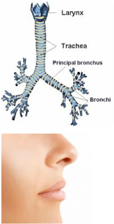
Larynx- The air then travels down past your larynx. The larynx or the voice box is located at the top of the trachea. The walls of the larynx are made of cartilage. Sound is produced by air passing through the larynx on the way to the lungs, causing the walls of the larynx to vibrate.
Trachea-After passing through the larynx the air makes its way through the trachea. The main function of the trachea is to allow air to pass in and out of the lungs. The trachea is a 12.5cm long tube made of cartilage. It is also referred to as the wind pipe. The trachea connects the voice box and bronchi and allows air to pass through the neck into the throat. Due to rings of strong cartilage the trachea remains permanently open.
Bronchi and Bronchioles- The main function of the bronchi and bronchioles is to transport air from the trachea into the lungs. On the end of the trachea the airways split into two bronchi braches. The left and right bronchi run into each respective lung before becoming smaller secondary bronchi. As the bronchi split into smaller and smaller branches they become bronchioles. The bronchi and bronchioles take air to the alveoli of the lungs. The bronchi use the cilia (tiny hairs that line the airways) to trap and move dust away from the lungs. During exercise the bronchi dilate which allows air to pass through the airways without resistance.
Cilia- Almost all of the airways, except for the mouth have fine hairs called cilia that are coated with a sticky mucus. The cilia trap dust and germs that enter your airways as you breathe. These particles are then swept up to the nose or mouth by the cilia where they're swallowed, coughed or sneezed out of the body.
Your airways are the pipes that carry oxygenated air to your lungs. They also carry carbon dioxide out of your lungs. The airways include your:
- Nose and mouth
- Larynx (voice box)
- Trachea (windpipe)
- Bronchi and bronchioles
Nose and mouth- The nose is the preferred entrance for air travelling into the respiratory systems. The nose filters the air coming in and regulates the temperature to ensure the oxygen can reach the lungs. The alternate entrance for air is through the mouth.
Larynx- The air then travels down past your larynx. The larynx or the voice box is located at the top of the trachea. The walls of the larynx are made of cartilage. Sound is produced by air passing through the larynx on the way to the lungs, causing the walls of the larynx to vibrate.
Trachea-After passing through the larynx the air makes its way through the trachea. The main function of the trachea is to allow air to pass in and out of the lungs. The trachea is a 12.5cm long tube made of cartilage. It is also referred to as the wind pipe. The trachea connects the voice box and bronchi and allows air to pass through the neck into the throat. Due to rings of strong cartilage the trachea remains permanently open.
Bronchi and Bronchioles- The main function of the bronchi and bronchioles is to transport air from the trachea into the lungs. On the end of the trachea the airways split into two bronchi braches. The left and right bronchi run into each respective lung before becoming smaller secondary bronchi. As the bronchi split into smaller and smaller branches they become bronchioles. The bronchi and bronchioles take air to the alveoli of the lungs. The bronchi use the cilia (tiny hairs that line the airways) to trap and move dust away from the lungs. During exercise the bronchi dilate which allows air to pass through the airways without resistance.
Cilia- Almost all of the airways, except for the mouth have fine hairs called cilia that are coated with a sticky mucus. The cilia trap dust and germs that enter your airways as you breathe. These particles are then swept up to the nose or mouth by the cilia where they're swallowed, coughed or sneezed out of the body.
Muscles Used for Breathing
Muscles near the lungs help expand and contract the lungs to allow for breathing. Some of these muscles include the:
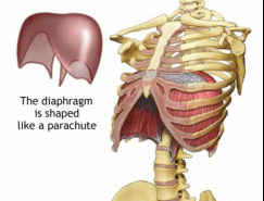
Diaphragm- The diaphragm is the main muscle in the respiratory system. It is a dome shaped muscles that is a key part in the breathing process. As you breathe in the muscles of the diaphragm pull downwards until they are completely flat which enclose the lungs in a protective cage. The diaphragm is an upward arching muscle.
Intercostal muscles- Intercostal muscles help to form and move the chest wall. They are found in between each rib. They expand and shrink the size of your chest cavity when you breathe in and contract when you breathe out.
Muscles near the lungs help expand and contract the lungs to allow for breathing. Some of these muscles include the:
- Diaphragm
- Intercostal muscles
Diaphragm- The diaphragm is the main muscle in the respiratory system. It is a dome shaped muscles that is a key part in the breathing process. As you breathe in the muscles of the diaphragm pull downwards until they are completely flat which enclose the lungs in a protective cage. The diaphragm is an upward arching muscle.
Intercostal muscles- Intercostal muscles help to form and move the chest wall. They are found in between each rib. They expand and shrink the size of your chest cavity when you breathe in and contract when you breathe out.
